DIAGNOSTIC IMAGING SERVICE
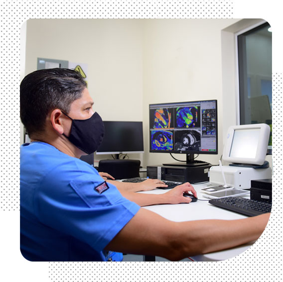
Diagnostic department
by images of the Omni Hospital is
committed to providing care
medical of the highest quality.
We put at your disposal specialist radiologists trained in the country and in prestigious centers abroad, which ensure better care. We have modern equipment that brings us up to the standard of current international medicine. Likewise, we have highly trained licensed personnel to execute the various imaging studies.
Our Diagnostic Imaging Services
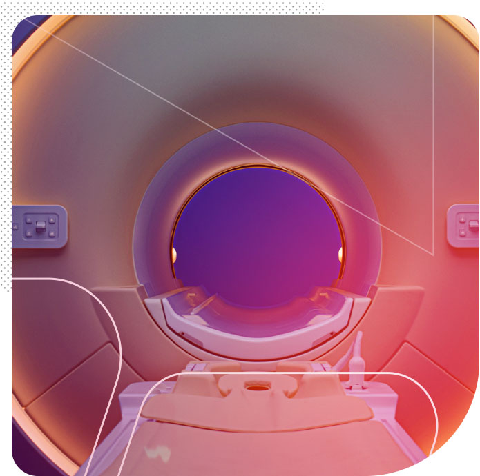
MRI is a study that uses magnets and radiofrequency waves to create images of the body, without the use of x-rays. It is one of the most recent technological advances in medicine for the accurate diagnosis of multiple diseases, still in the early stages. It is made up of a complex set of electromagnetism emitting devices, radio frequency receiving antennas and computers, which analyze data to produce detailed images, two or three dimensions with a high level of precision, which allows creating images of the body, without the use of X-ray and thus be able to rule out alterations in the organs and tissues of the human body.
Magnetic Resonance is ideal for pediatric application thanks to the absence of ionizing radiation, being a safe, non-invasive study with no known side effects.
At Omni Hospital we have a sedation and/or anesthesia service to offer solutions to claustrophobic and pediatric patients.
We have specialized equipment and trained personnel to safely perform this type of procedure within a magnetic field.
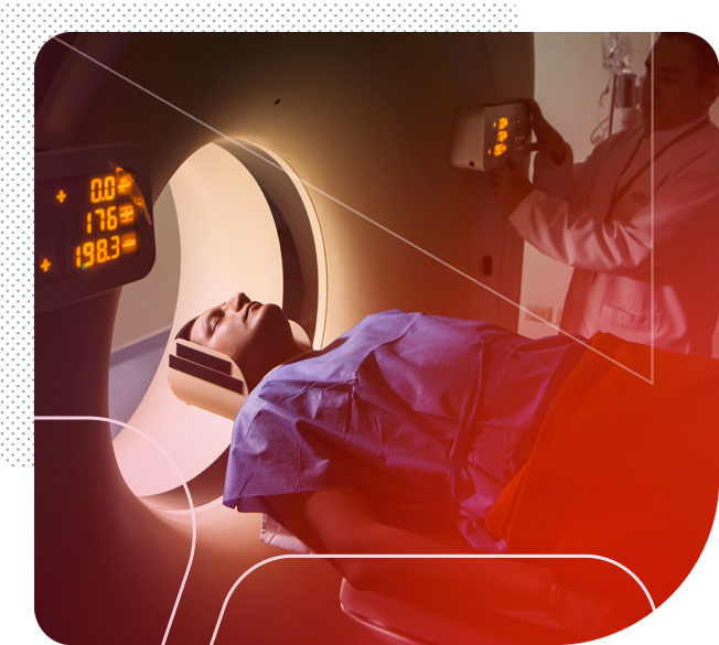
Multislice tomography is a study that uses ionizing radiation and an advanced computer system to quickly and accurately obtain high-resolution images of both soft tissues and bone structures.
The images show with high clarity various tissues such as the liver, spleen, pancreas, kidneys, among others; and they allow small lesions to be analyzed and differentiated into benign or malignant processes, congenital malformations or trauma.
It is an ideal procedure for:
• Analyze small lesions and differentiate them into benign or malignant processes.
• Perform biopsies and drain abscesses.
• Create 3D images of bone structures, urinary tract (urotc), respiratory (virtual bronchoscopy), abdominal (virtual colonoscopy) and blood (angiotc).
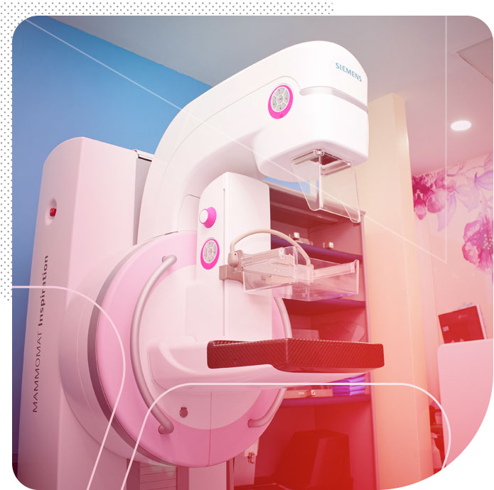
From the age of 40 it is recommended that all women have a control mammogram every year. If there is a history of breast or ovarian cancer in your family, it is advisable to start check-ups from the age of 30.
At OmniHospital we have the best technology to carry out studies such as: 3D mammography with tomosynthesis which detects more types of cancer than a standard 2D mammography, since with a three-dimensional image the disease can be detected in people without symptoms, reducing the rate of false positives.
Three-dimensional mammography is becoming more common, but it is not available in all centers, so when you do your medical exam, do it with the best equipment in the best place.
Mammography is the radiological study of the mammary gland, that is, an X-ray of the breasts. It is the only tool that allows accurate diagnosis of breast carcinoma in early stages when it is not palpable, for this reason it is the only method used and approved worldwide in the investigation of the disease.
Services:
• 2D digital mammography.
• 3D digital mammography Tomosynthesis.
• 2D digital mammography plus Tomosynthesis.
Procedures with Sterotaxia:
• For pre-surgical harpoon placement.
• Minimally invasive biopsies with Sterotaxica guide
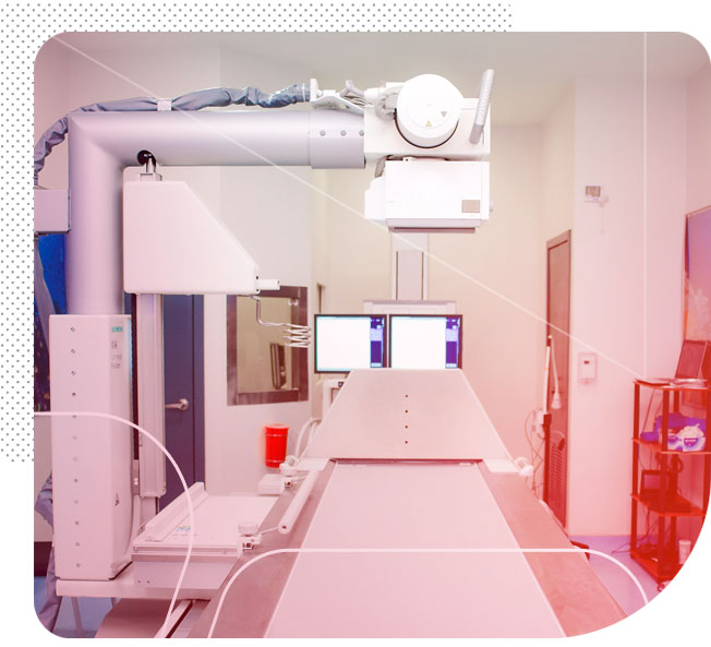
An X-ray is a study that uses X-rays, which are electromagnetic waves that can pass through any anatomical region and produce images of the interior of the body.
The x-rays are absorbed by the part of the body to be studied and generate an image on the radiographic plate or cassette.
X-rays show the anatomy of the human body in gray scales. They are especially useful in bone and joint evaluations. They are also essential in cardiovascular and pulmonary studies.
Thanks to our digital radiology equipment we achieve a lower radiation dose to the patient, with greater efficiency and flexibility.
Thanks to our digital radiology equipment we achieve a lower radiation dose to the patient, with greater efficiency and flexibility.
There are simple x-ray studies such as: chest, paranasal sinuses, extremities, among others; and also special contrasted studies that require the intervention of the radiologist and the administration of a contrast medium such as: Esophagogram, intestinal transit, colon by enema, voiding cystography, hysterosalpingography, sialography, among others.

Echocardiography is a cardiac exploration technique that allows the structural and functional study of the heart through the medical application of a physical phenomenon, such as ultrasound. It is a fundamental exploration method for the diagnosis of heart patients or those with suspected cardiovascular disease.
This type of ultrasound provides information about the shape, size, function, and strength of the heart. It also evaluates the thickness of the walls and the operation of its valves.
Stress echocardiogram It is a non-invasive test, with which the movement of the heart can be evaluated when it is at rest and compared with the result obtained after the administration of a drug called Dobutamine. This medicine causes the heart to beat rapidly so that it can determine if there is decreased blood flow through the coronary arteries.
Transesophageal echocardiogram It is a moderately invasive study in which a probe or tube is inserted through the mouth, reaching the esophagus, which allows the specialist to see, more closely, the functioning of your heart (cavities, blood flow, valves).
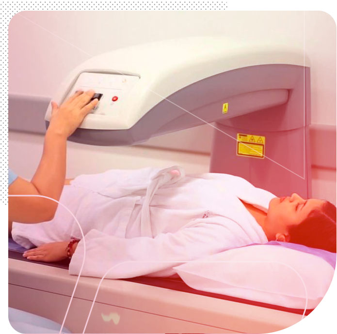
Bone densitometry is a diagnostic method used to study bone tissue with which the degree of bone calcification is determined using very low doses of radiation. It is performed on the hips, lumbar spine, forearms and full body.
Its precision, accuracy and reliability are based on easy and fast performance with minimal exposure to radiation and easy interpretation of the results.
Its main indication is the suspicion of osteoporosis, a systemic skeletal condition where there is a decrease in bone mass, as well as a deterioration of the microarchitecture of bone tissue and, therefore, greater bone fragility and risk of fractures. All of this will allow your treating physician to obtain an accurate diagnosis of osteopenia or osteoporosis, in order to initiate specific treatment, in addition to having a record and monitoring of the disease.
Bone Densitometry also allows the determination of body fat content and other important parameters for the follow-up and control of patients under treatment with anabolic steroids, growth hormones, weight control medication, among others.
AVAILABLE AT AVALON PLAZA III OMNIHOSPITAL SPECIALTY CENTER.
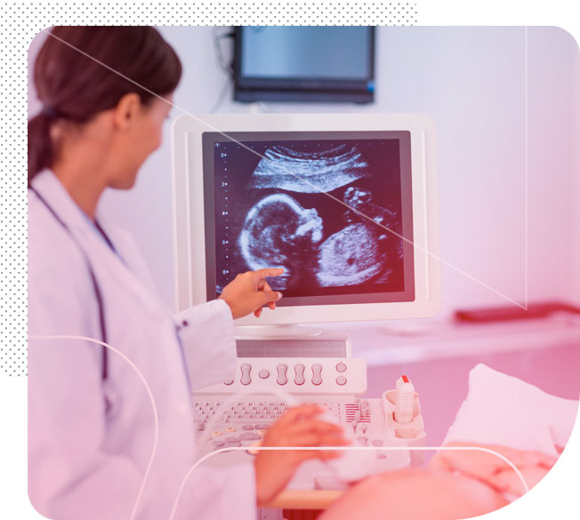
Ultrasound is a study that produces images of the interior of the body in real time, by recording the echoes emitted by the different tissues of the anatomical area explored when they receive vibrations or sound waves from the transducer.
It is a safe, non-invasive, painless and easy-to-perform study, achieving greater diagnostic reliability given the quality of the images obtained. It also allows directing the placement of catheters for drainage of collections and the performance of ultrasound-guided biopsies of suspicious breast, prostate, thyroid and liver lesions.
Diagnostic imaging service
The diagnostic imaging department at Omni Hospital is committed to providing the highest quality medical care. We put at your disposal specialist radiologists trained in the country and in prestigious centers abroad, which ensure better care. We have modern equipment that puts us up to the standard of international medicine imaging studies.
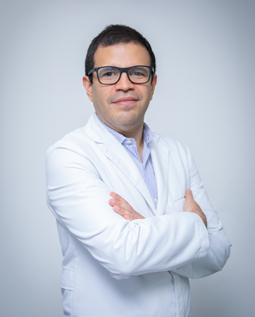
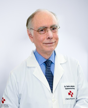
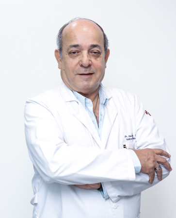
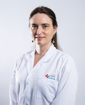
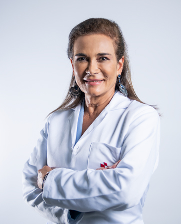
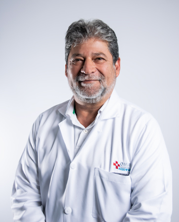
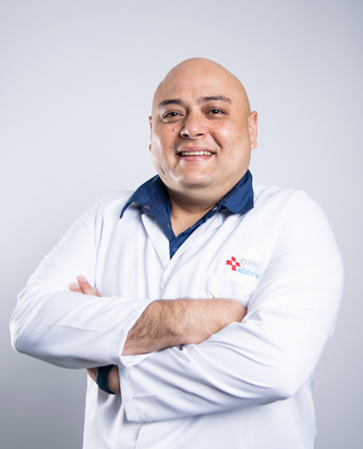
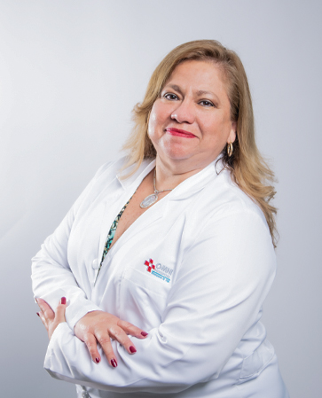
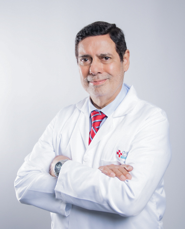
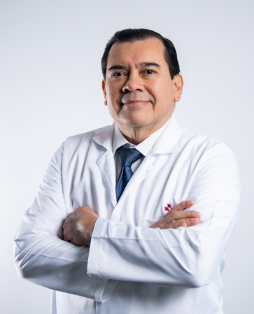
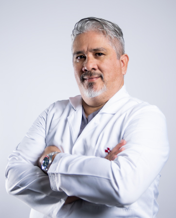
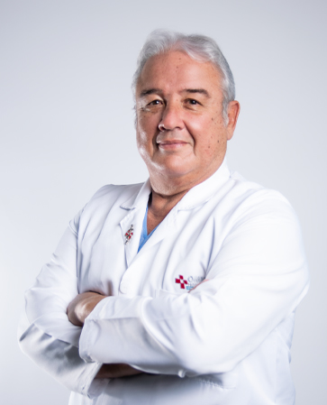
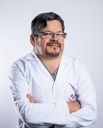
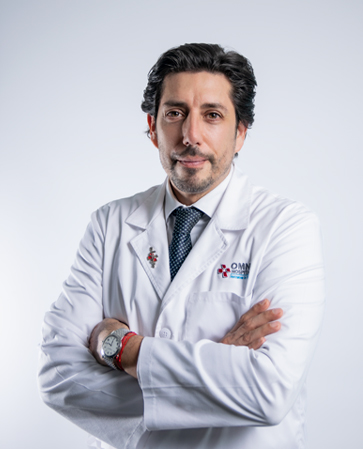
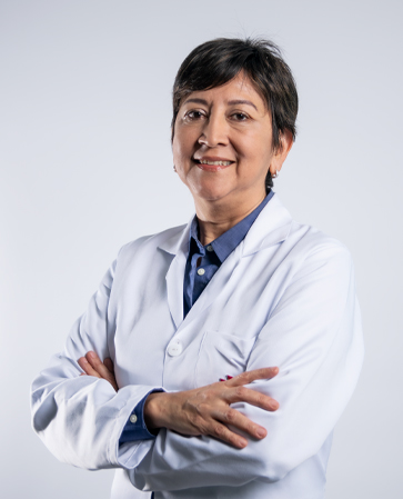
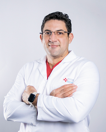
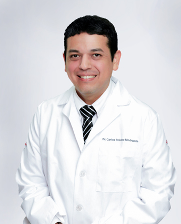
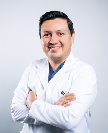
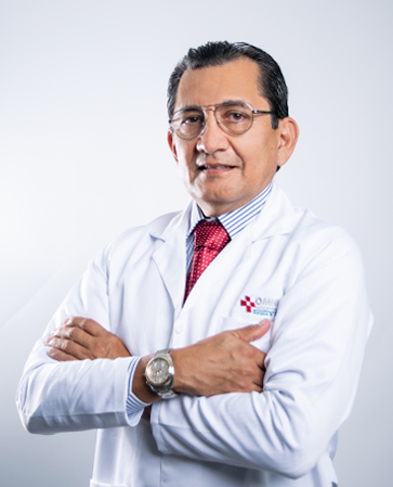
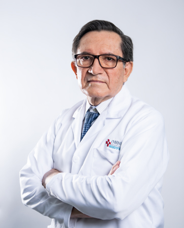
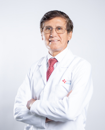
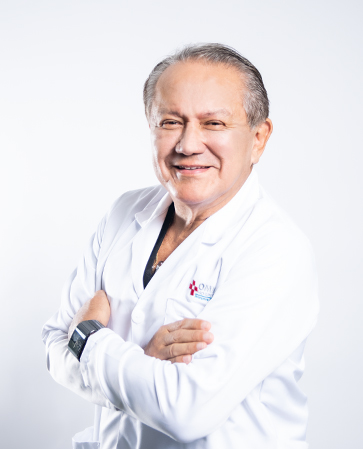
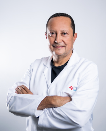
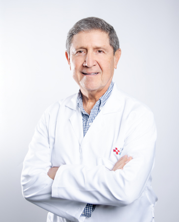
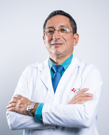
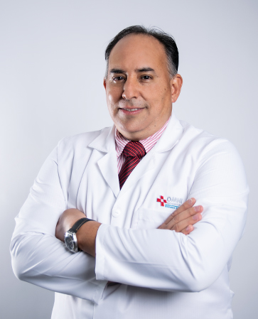
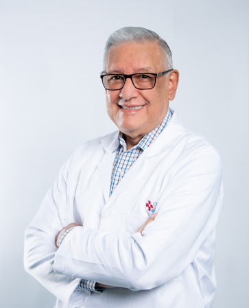
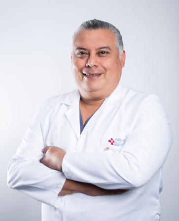
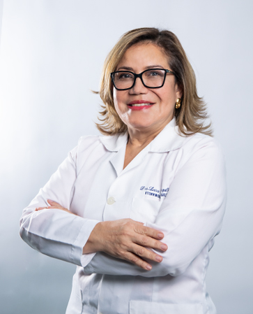
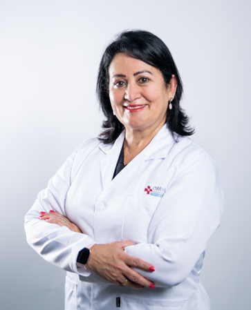
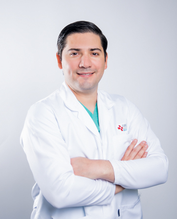
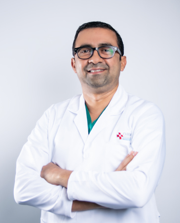
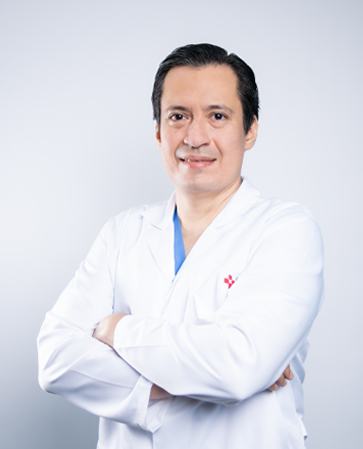
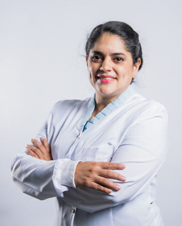
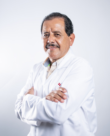
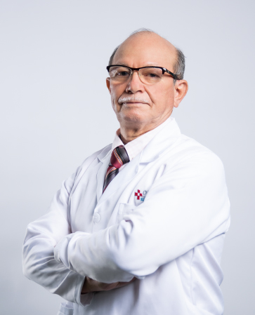
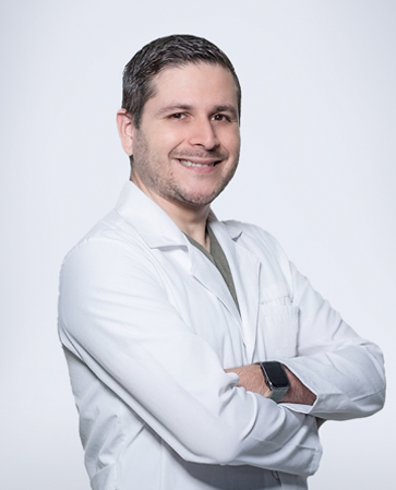
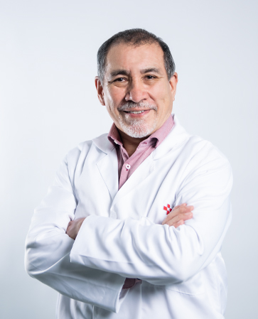
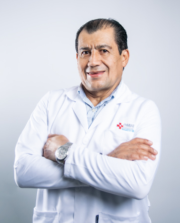
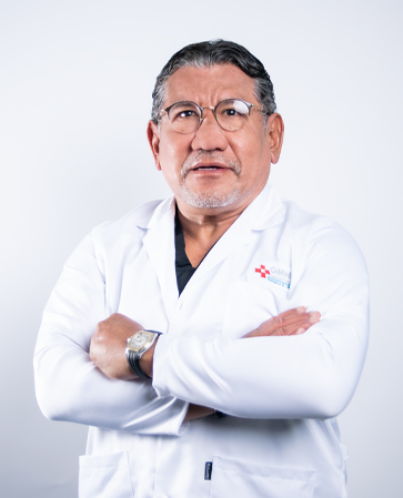
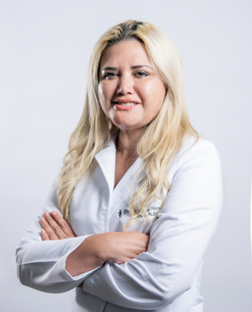
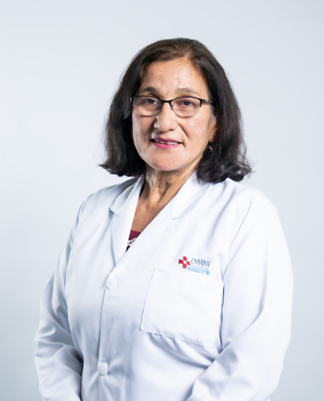
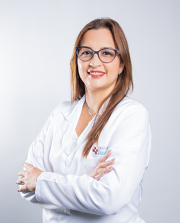
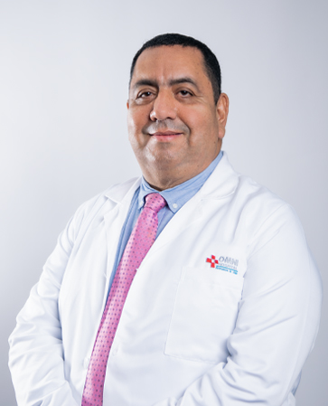
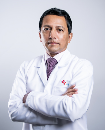
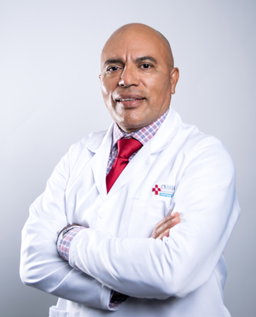
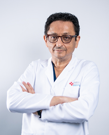
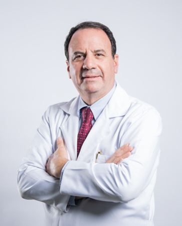
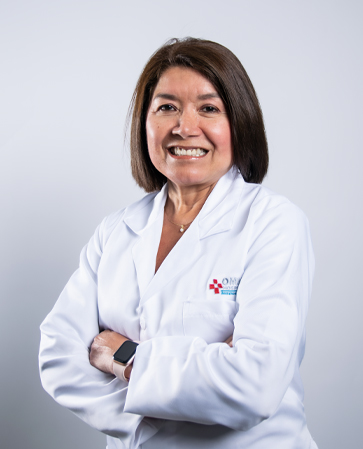
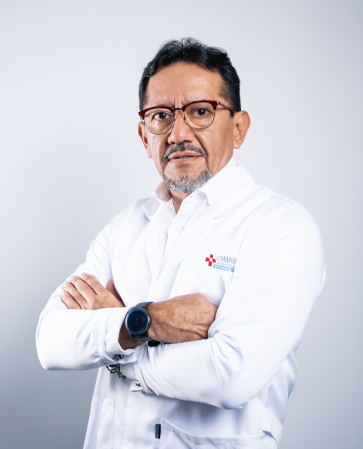
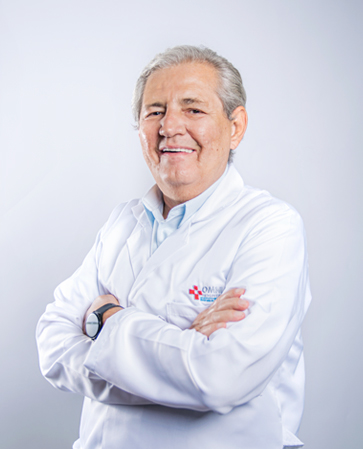
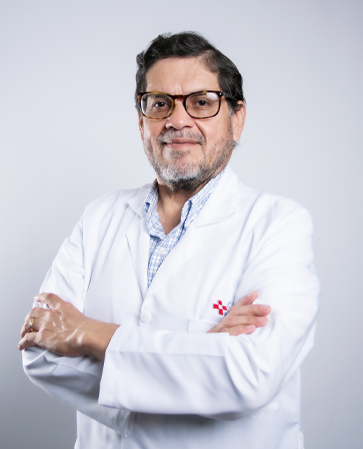
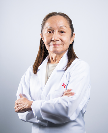
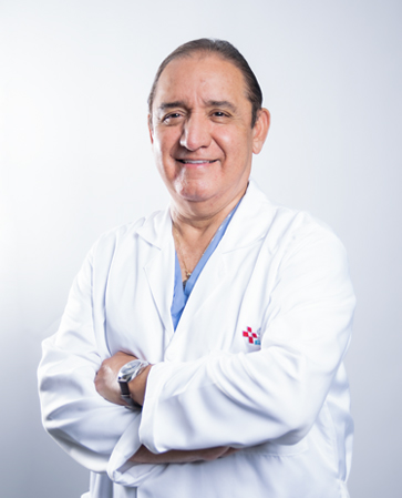
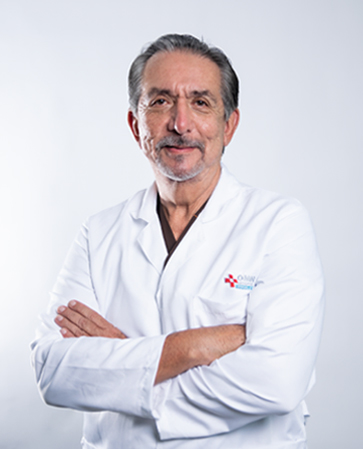
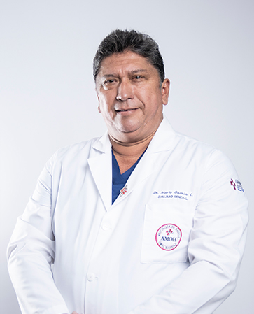
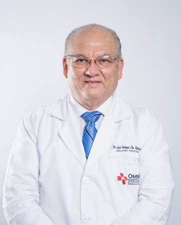
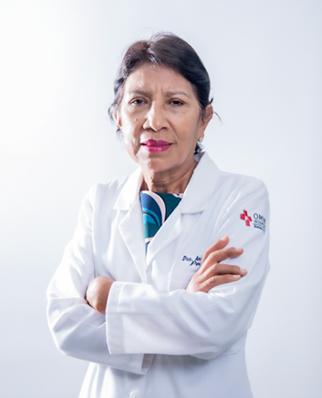
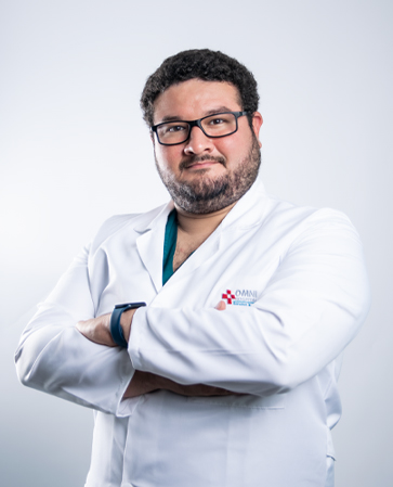
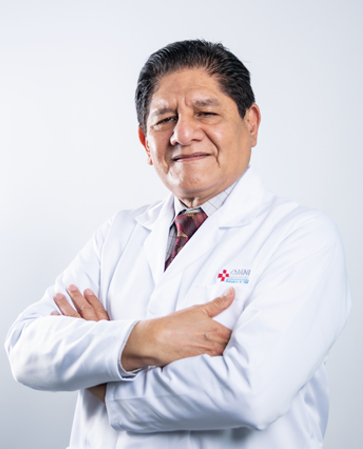
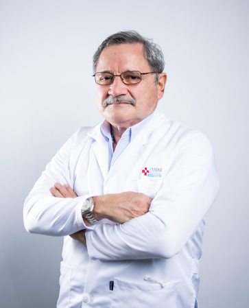
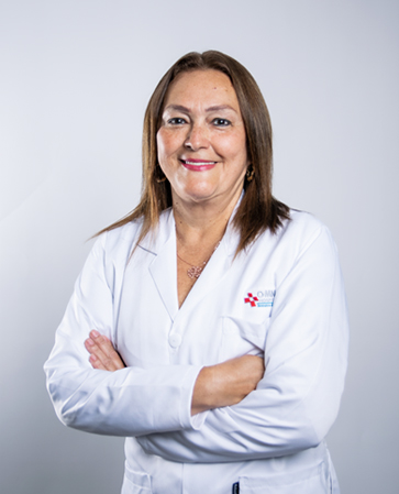
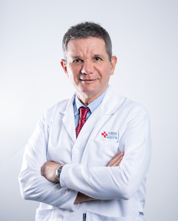
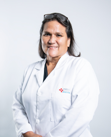
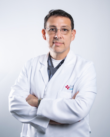
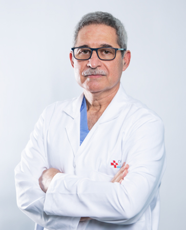
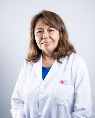
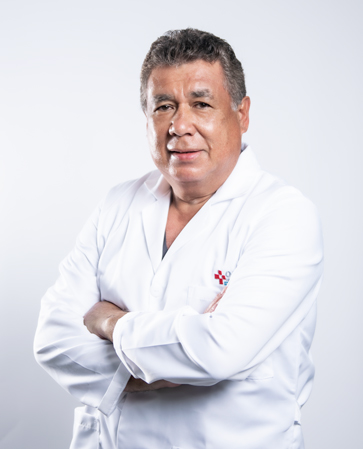
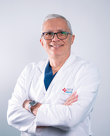
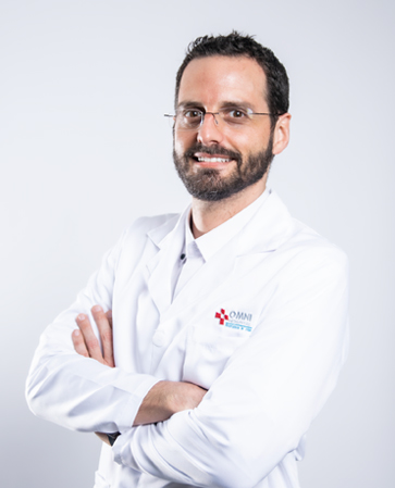
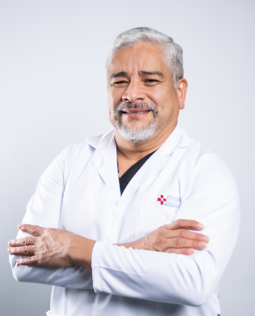
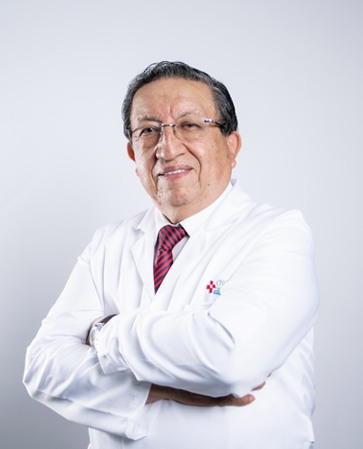
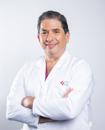
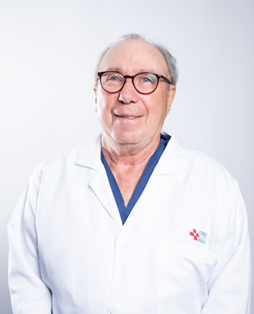
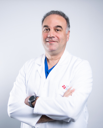
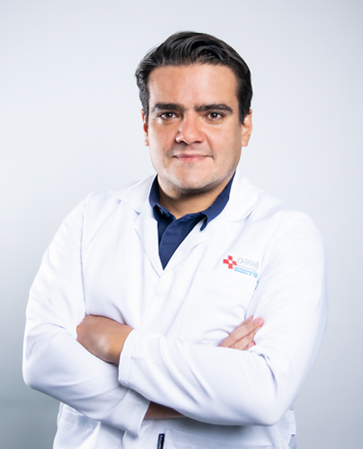
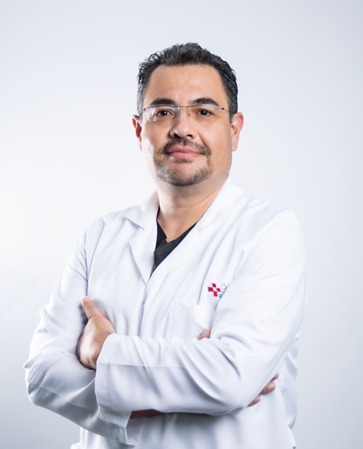
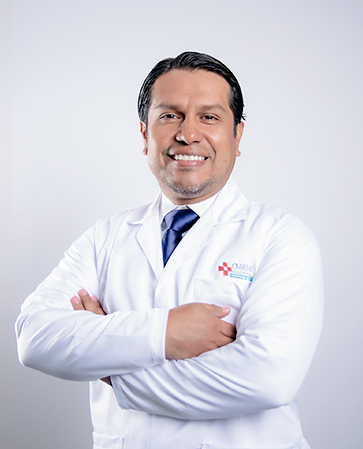
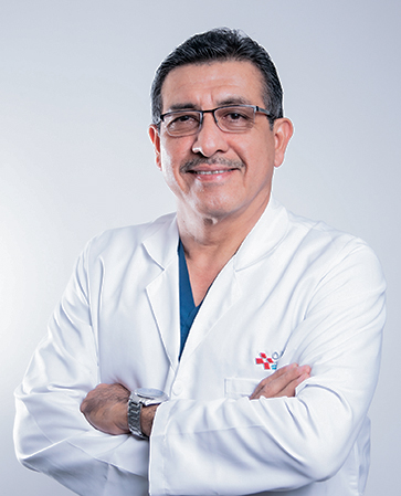
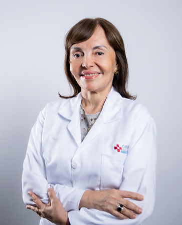
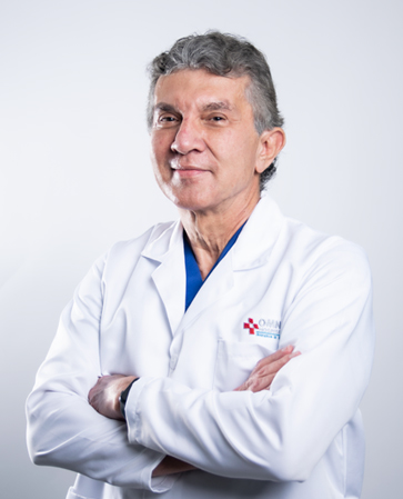
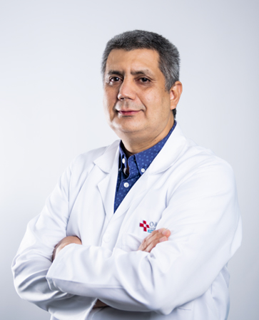
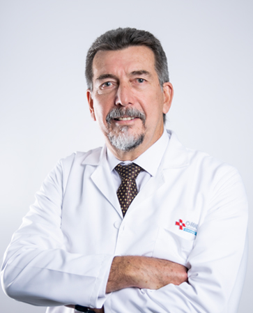
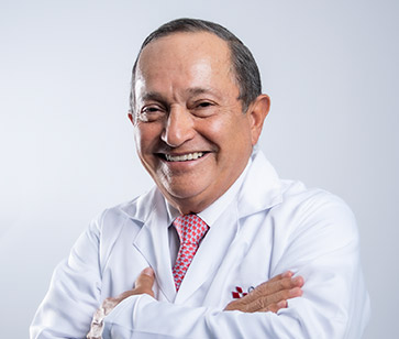
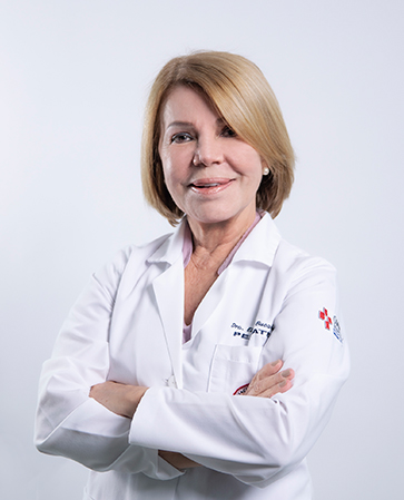
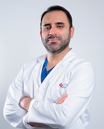
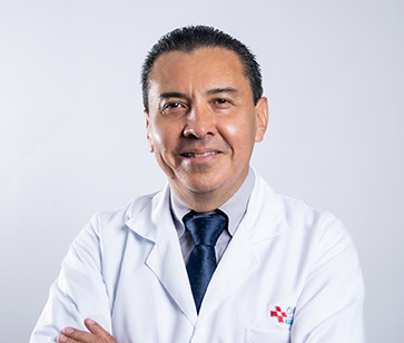
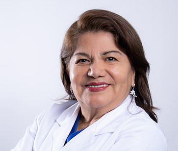
Radiologists

Dr. Ángel Alvarado Lema

Dra. Isabel Faria Urdaneta
Dra. Alicia González Guarnizo

Dr. Christiam Palacios
Médicos Ecocardiografístas

Dra. Cruz Simigliani Hernández

Dr. José Dávila Terreros

Dr. Ricardo Rosales Ramos
Adults

Dra. Elsie Valdivieso Valenzuela

Dr. Freddy Balda Caravedo

Dr. Eric Macías Ochoa

Dr. Joel Moreno Uzcátegui

Dr. Stewart Blum Astudillo
Pediatric

Dra. Ileana Gómez Rojas
Dr. Simón Duque Solórzano

Dra. Patricia Árias Alarcón


